
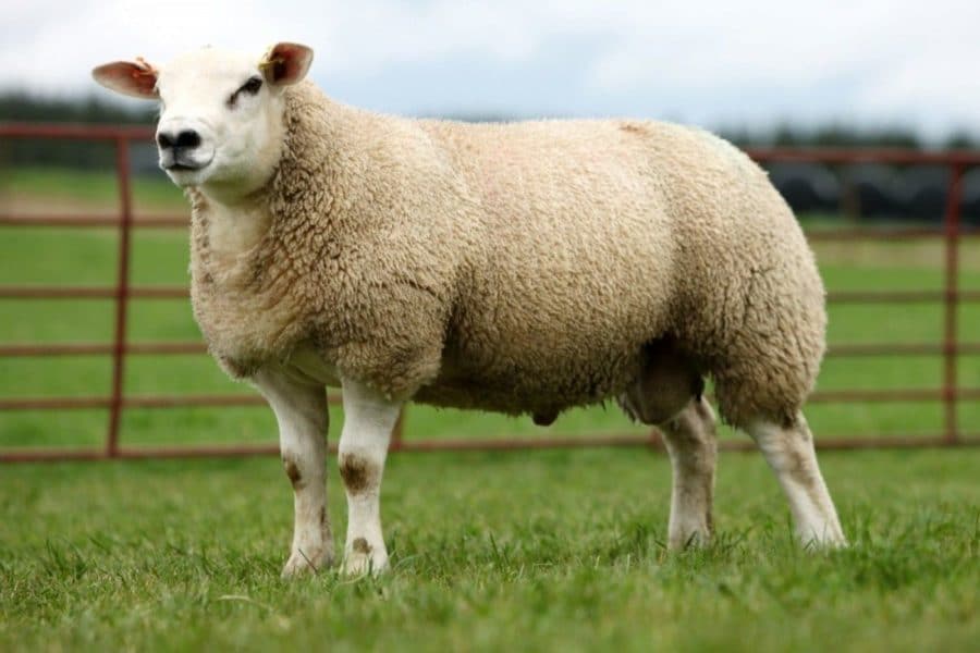
Texel Sheep: Characteristics, Meat Quality and Production
April 6, 2026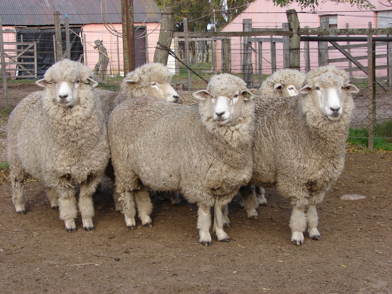
Corriedale Sheep: Characteristics, Wool, Meat and Production
April 6, 2026Actinomycosis, commonly known as lumpy jaw, is a chronic bacterial infection that causes progressive, hard swellings in the jaw, face and occasionally other body parts of sheep and goats. While not immediately fatal, it significantly affects the animal’s ability to eat, leading to progressive weight loss and, if untreated, death from starvation.

What Is Actinomycosis?
Actinomycosis is caused by the bacterium Actinomyces bovis (and related species). It is a gram-positive, anaerobic filamentous bacterium that is normally present in the mouth and upper respiratory tract of ruminants as a commensal organism.
Infection occurs when the bacterium gains entry through damaged oral mucosa — typically caused by:
- Injuries from rough, sharp feedstuffs (stalks, dry hay, grain awns, thistles)
- Dental eruption wounds (young animals)
- Tooth root infections
- Oral lacerations from foreign objects
Once established in the bone tissue of the mandible (lower jaw) or maxilla (upper jaw), the bacteria cause a chronic granulomatous osteomyelitis — a slow, progressive destruction of bone tissue with formation of characteristic lesions.
Symptoms of Actinomycosis in Sheep and Goats
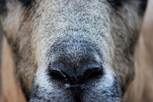
- Hard, painless swelling on the lower jaw (mandible) — the most characteristic sign. The swelling is bony-hard to the touch and does not move.
- The swelling grows slowly over weeks to months
- Later stages: the swelling may soften in the center and rupture, releasing a thick, honey-colored pus containing characteristic “sulfur granules” (yellow-white granules visible to the naked eye)
- Difficulty chewing and eating — the animal shows progressive preference for soft foods, reduced feed intake, quidding (dropping partially chewed food)
- Progressive weight loss and poor body condition despite available feed
- Tooth loss or loosening in advanced cases
- Drooling and bad breath
Note: Do not confuse actinomycosis with caseous lymphadenitis (CLA), which also causes swellings in sheep and goats but affects the lymph nodes rather than the bone, and is caused by a different bacterium (Corynebacterium pseudotuberculosis). CLA abscesses are softer and found at specific lymph node locations (jaw angle, flank, shoulder), while actinomycosis affects the bone of the jaw itself.
Diagnosis
Diagnosis is primarily clinical — based on the characteristic hard jaw swelling, history and physical exam findings. Confirmation can be obtained by:
- Pus examination: Presence of sulfur granules in discharged material is highly diagnostic
- Bacterial culture and sensitivity: Confirms species and guides antibiotic selection
- Radiography: Shows characteristic bone destruction (lytic lesions) and new bone formation
- Histopathology: Shows granulomatous tissue with characteristic actinomyces colonies (“ray fungus” appearance)
Treatment of Actinomycosis
Treatment is most effective in early stages, before extensive bone destruction has occurred. Options include:
Antibiotic Therapy
- Penicillin: First-line treatment. High-dose, long-duration therapy required (3–6 weeks). Intramuscular or intravenous administration.
- Tetracyclines: Oxytetracycline or doxycycline — effective alternative, particularly useful for long-term oral administration.
- Trimethoprim-sulfonamide combinations: Used when penicillin is unavailable.
Iodine Therapy
Oral sodium iodide (20 mg/kg IV or 1 g sodium iodide per 15 kg body weight orally for 30 days) has been used traditionally and is still considered a useful adjunct treatment. Iodides help penetrate bone and fibrous tissue where antibiotics have poor access. Signs of iodine toxicity (excess tearing, nasal discharge, coat flaking) indicate need to reduce dose.
Surgical Drainage
In cases where the swelling has softened and fluctuating pockets of pus have formed, careful surgical drainage followed by irrigation with iodine solution or hydrogen peroxide can help. However, surgery alone is not curative — must be combined with antibiotic treatment.
Prognosis and When to Cull
Prognosis depends heavily on how early treatment is initiated:
- Early-stage cases: Good response to treatment; bone lesions may stabilize
- Advanced cases with extensive bone destruction: Poor prognosis — treatment may slow progression but rarely achieves complete cure
- Animals that cannot maintain body condition despite treatment: Culling is the most humane and economically sensible decision
The cost of long-duration antibiotic treatment must be weighed against the animal’s value. For high-value breeding animals, aggressive early treatment is warranted. For commercial flock animals, early culling before significant weight loss is often the most practical decision.
Prevention
- Feed management: Reduce exposure to sharp, abrasive feeds (hay with sharp awns, thistles, dry stalks). Chop or soften feedstuffs for older animals with dental wear.
- Biosecurity: Quarantine new animals and inspect jaw and lymph nodes before introducing to the flock
- Regular dental inspection: Detect broken teeth, tooth root problems and early-stage swellings during routine handling
- Avoid overcrowding: Reduces competition for feed and associated oral injuries from fighting
- Good pasture management: Maintain pasture at a height that prevents animals from ingesting soil and debris that may carry bacteria
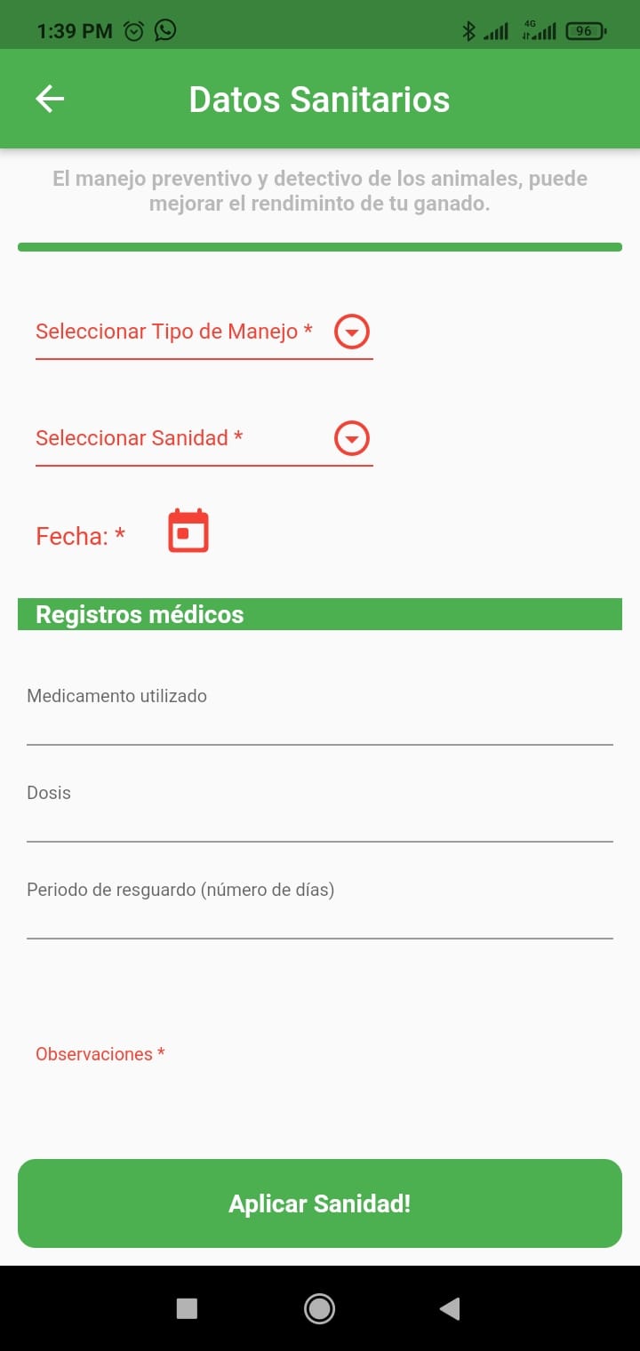
Recording health events and treatment responses for each animal in OvinApp allows you to track cases of actinomycosis in your flock, evaluate treatment outcomes and make better culling decisions based on individual animal history rather than guesswork.